|
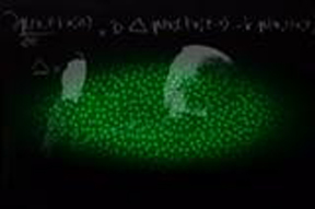
Time-lapse movie of drosophila embryonic development
Video and text created by Jean-Baptiste Boule, Matthieu Coppey, and
Thomas G of www.princeton.edu and posted in Google on May 13,
2006.

Click image to play video (52sec)
Recent advances in microscopy and in fluorescence labeling of proteins
allow the observation of complex biological processes in living
organisms. This movie shows the early stages of embryonic development
of a fruit fly egg, which is half a millimeter in size. In the first part of the
movie, the observation of fluorescently labeled histones (proteins that
help packaging the chromosomes into a compact nucleoprotein structure in the cell) reveals the dynamics of the mitotic divisions of the nuclei that
occur in the egg during the first two hours of development. Following the
synchronous nuclear divisions, cellular membranes form and cell movements initiate a process called gastrulation, during which cells
invaginate in the embryo to form tissue layers and organs following a
genetically determined developmental program. Early steps of
gastrulation are shown in the second part of the movie through
fluorescence labeling of membrane proteins. As suggested in the movie by the biophysicist writing down differential reaction-diffusion equations
on a blackboard, the ability to observe these kinds of processes at the
molecular level and in real time has opened new venues in modern
biology, by applying tools from mathematics and physics to understand complex biological processes in a quantitative manner.
----------------------------------------------------------------------------
Click here for previously featured Science links of the week
|